The future is dark, which is the best thing the future can be, I think (Virginia Woolf)
It is only just before 7 and the sun has not yet decided to rise. I am at a teaching hospital where I am meeting Amir, a radiologist, who has agreed to let me observe him at work. We take the stairs down to the basement and make our way to the radiology department. I see all the hospital gowns neatly put in stacks on one of the tables, waiting for the patients. We pass the scan rooms with shielded walls and windows to protect from radiation exposure, holding all the technological equipment needed to take medical images. Then we reach the reading room, which is a dark and quiet room filled with computer screens. When the radiologists arrive in the morning, they have been assigned to read specific images. If their lucky, Amir says, they can manage to read the images of around 20 patients during a day. The radiologists follow the list chronologically. They look at the images, scroll up and down, zoom into images, and talk quietly, more like whispering, into dictation devices. Despite the Alberta Bulletin’s predictions from 1962 that radiologists would come out of the darkness and into the light, here we are (Hall, 1962). In the darkness. Like moths the radiologists are drawn to the light of the screens. They need breaks sometimes to rub their eyes. Eventually, they adapt to this darkness they say. I soon learn that a large part of a radiologist’s work includes looking for shadows. Shadows that do not look quite right. To a novice like me there are hundreds if not thousands of shadows in an image and I cannot tell if a specific shadow looks suspicious, not even when Amir points them out to me. What I have learned, however, are some methodological lessons. Or rather, some counter lessons.
Lesson 1: Darkness & Contrast
Darkness is a condition that helps radiologists read medical images. They turn down the lights to see better. The dark reading room increase image contrast and the various shade of grays becomes clearer when the lights are turned down. Radiologists need that contrast. The shades of gray the human eye can perceive depends on these conditions. In a way, radiologists use darkness to improve their vision. To enhance their ability to interpret images accurately. To find the smallest details and detect subtle abnormalities. The darkness also helps the radiologist to focus on the images and not get distracted from their surrounding environment. Darkness improves their possibility to do their work well. This is the first lesson – in radiology practice darkness and contrast is a valued condition.
Lesson 2: Uncertainty
As Amir is scrolling up and down the images, he finds something that worries him. “See, this little guy here” he says, pointing to one shadow among hundreds of others. “This does not look right to me”. He measures the shadow and marks it on the image so that others looking at the images can find the shadow that he is referring to. He puts the measurements in the image as well. The shadow that he found is 6 mm long. He talks into his dictation device quietly, reporting the shadow that will now end up in the patient information. He mentions the dimensions of the shadow and the number of the image where the mark he has made can be found. A shadow that he refers to as “a lesion”. “When I do not know what it is, I call it a lesion”, Amir says. He explains that the patient’s doctor needs to take a closer look at this shadow and decide the next step. The patient might need to do further scans or tests to decide if this is something to worry about or not. The uncertainty of what his findings might be does not bother him. He looks for shadows that does not look quite right, mark them in the image, and make a note about it. This needs to be explored further. This then, is the second lesson – radiologists do not shy away from uncertainty.
Lesson 3: Change
Sometimes the patient has had medical images taken before of the same area of the body as they have taken images now. Amir says that this is good because then they can look at older images and compare. This happens when Amir and I look through the images of a patient. Amir notices an abnormality. He goes through the patient’s information and find that the patient has had medical images taken 8 years ago. If the patients had medical images from before, Amir can go back and take a closer look to see if the shadow he now found was there then as well. He opens the older images next to the newer ones. He tries to find the area in the older picture where the shadow that worries him should be if it was there before. He scrolls up and down and looks carefully at the images. He zooms in and out and look at the area from different perspectives. After a while he says that he can find the shadow in the older images. The shadow looks exactly the same today as it did 8 years ago, and therefore it is probably nothing to worry about. He still marks the shadow and talks into his dictation device. He adds that he has been looking at the older images available and that the shadow was present then as well. Then the patients doctor decides what should happen next, Amir says. It is really good when patients have previous images, of the same area, taken before because then we can compare. We look for change, Amir says. Has anything changed? Is anything different? This is the third lesson – look for change.
Lesson 4: Look Beyond
What’s the clinical problem? That is the first thing we look for, Amir teaches me. We first look through the patient information. Here we can see the notes that the patient’s doctor has written. When medical images are taken, there is always a clinical question, says Amir. “However,” Amir says, “we as radiologist have to go through every organ carefully to look for abnormalities. The thing is – about 30 % of the images we go through show something different than what we are looking for. That is, something different than the clinical question. We find other things than the patient’s doctor specifically asked us to look for”. This happens to be the case with this patient. Amir takes his time and looks at every organ in detail. Let’s take a closer look at our next patient. Amir reads the patient information and tells me that the clinical questions is: does this patient have kidney stones. Amir continues, “we of course look for kidney stones and as you can see the patient’s kidneys look just fine”. Amir goes on and tells me what it would have looked like if the patient had kidney stones. Even I can see the evenness in the shadow we are looking at that represents the kidneys. The answer to the clinical question is “no, this patient does not have kidney stones”. After a while he says, “I found something. Look at this thing here. Do you see that? Here on the spine.” In an otherwise even shadow, I too can spot a small difference. The radiologists go in with a particular clinical question, in this case: does the patient have kidney stones. The answer is: no. However, the radiologists do not let this restrict them. They look for other things as well. The fourth lesson is: radiologists go beyond the question at hand.
Dark Methods as Thought Experiment, or the value of Counter Lessons
Having discussed some lessons learned from radiology, how can such insights inform theoretical thinking and guide fieldwork? I think that we can learn something from how radiologists work with ‘darkness and contrast’, ‘uncertainty’, ‘change’, and ‘going beyond the question at hand’. In radiology, these are valued conditions and examples of how darkness becomes a tool that they work with. Feyerabend ([1975] 2010) talks about counterinduction, that is, hypothesis that are used to effect change in its opposite. Hypothesis that contradicts already existing and established theories and observational results. Feyerabend ([1975] 2010) argues that we might advance science by working counterinductively. What’s at stake in Feyerabend’s proposition is challenging assumed principles. We are often not even aware of the assumptions we make. How can we make them visible? How can ideas such as counterinduction be worked with in practice?
We know that in disciplines, it becomes custom to think about something and explore something in a specific, established way. What we need is something that helps question how something is commonly described. Something that can help us contradict what is already established and not fall into what we assume to be the most plausible principle, what is most likely, responses that we are accustomed to. Something that will help us think differently and interfere with the commonly accepted. What if we were to value the idea of getting lost a bit more and allow ourselves to go a little feral? Exploring the uncertain in terms not just of empirics, but also with ways we know. What if we alongside what is commonly known or established practice in research also valued the experience of following the empirics more than our hypothesis? Going beyond what and how we know. Practicing an openness towards alternative understandings.
What kind of figure is darkness? The attributes of darkness are not typically related to seeing better. In Western philosophy and thinking, darkness comes in binary relations with light, generally understood as the negation of any characteristics of light (Dunn and Edensor, 2020). But what if we engage with darkness as a positive, as an enabler, like our friends in radiology do? Darkness as a way to work with contradictions, with the unexpected, with the counterintuitive. Engaging with darkness as a thought experiment of thinking in reverse? Keeping the idea that it could be otherwise (Woolgar and Lezaun, 2013). Returning to radiology, darkness and contrast, in uncertainty and change, and by going beyond holds great potential when it comes to dispute established knowledge, encourage new observations, and new ways of doing and thinking about things. From our friends in radiology, we learned that darkness and contrast could be used as ways to make things more clearly. A tool to improve vision. Radiologists were not bothered by uncertainty. Instead, they worked with change and went beyond the question at hand, exploring other things as well. Darkness as a way to learn to expect the unexpected. Working with such tools, enriched their practice. In this way, darkness could do important ideological work, challenging and interfering with what is established. An invitation to explore alternative understandings, opening doors toward other worlds, making other things possible. Darkness as a counter movement. Darkness, as what Solnit (2006) would call a field guide to getting lost.
It is time for me to go. I take the stairs and walk back up to the entrance and leave the hospital. The radiologists, however, still have a few hours of work left. The radiologists, then, will stay in the dark.
‘Let it be dark’ (Ursula Le Guin)
Referenser
- Dunn, N and Edensor, T (2020) Rethinking Darkness. Cultures, Histories, Practices. London: Routledge.
Feyerabend, P ([1975] 2010) Against method. London & New York: Verso. - Hall, R. M (1962) From Darkness Into the Light. Alberta Medical Bulletin 27(2): 56-60.
- Le Guin, U (1969) Left Hand of Darkness. New York: Ace Books.
- Solnit, R (2006) A Field Guide to Getting Lost. London: Penguin Books.
- Woolf, V (1979) The Diary of Virginia Woolf 1915-19. Boston: Mariner Books.
- Woolgar, S and Lezaun, J (2013) The wrong bin bag: A turn to ontology in science and technology studies? Social studies of science 43(3): 321-340.
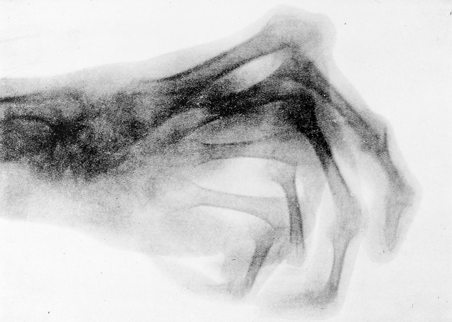
Illustration: Röntgenvågor i medicinskt arbete (1907) av David Walsh. Wellcome Collection.
